Long Bone Diagram Endosteum / Bones Anatomy Physiology Wikivet English / Cortical bone appears radiopaque (white) on radiographs as the outermost layer of bone.
Long Bone Diagram Endosteum / Bones Anatomy Physiology Wikivet English / Cortical bone appears radiopaque (white) on radiographs as the outermost layer of bone.. The diaphysis and the epiphysis. Osteocytes synthesize bone and reside on the surfaces of bone: Flat bones, like those of the cranium, consist of a layer of diploë (spongy bone), lined on either side by a layer of compact bone ().the two layers of compact bone and the interior spongy bone work together. Types of bone ossification 6. The inferior end.,anatomy of a long bone ms.
It is found in bones such as the humerus and the. Give your diagram a caption or heading. Make sure that you follow all the guidelines for biological drawings: A long bone has diaphyseal bone is organized to create the best balance between weight and structural strength. The endosteum appears at the interface between the.
Notes Ch 7 Skeleton from www.biologycorner.com The endosteum appears at the interface between the. Compact bone that forms the shafts of long bone consists of two structures. Osteocytes synthesize bone and reside on the surfaces of bone: They include the clavicle, humerus, radius, ulna, femur, tibia, and the inner surface of compact bone is lined by a thin, cellular layer, the endosteum. • the sections are then cut and stained with hx and eosin to • the long and short hones are formed externally of compact bone, but their endosteums are irregular due to presence of spongy bone. Long bones are those that are longer than they are wide. Newly formed bone originating from endosteum was observed on day 6. The ossification/bone formation occurs either as endochondral or as intramembranous osteogenesis.the difference lies in the presence of bone formation:
It is found in bones such as the humerus and the.
Compact bone that forms the shafts of long bone consists of two structures. Types of bone ossification 6. The delicate connective tissue layer lining the inside surface of compact bone. Endosteum, centrally, which is derived from derived from a condensation of inner connective tissue, and which helps separate the marrow cavity. General concepts about skeleton 2. A long bone has diaphyseal bone is organized to create the best balance between weight and structural strength. The endosteum is located on the internal surface of the bone, being the membranous layer that covers the medullary cavity, the bony trabeculae (spongy part of the bone), the haversian canals and internal walls of the compact long bones. Bones are treated with nitric acid to remove their calcium. Cortical bone appears radiopaque (white) on radiographs as the outermost layer of bone. Bone as an organ 3. At the ends of the bone the periosteum is continuous with the joint. Both the periosteum and the. The endosteum (plural endostea) is a thin vascular membrane of connective tissue that lines the inner surface of the bony tissue that forms the medullary cavity of long bones.
Types of bone ossification 6. Compact bone that forms the shafts of long bone consists of two structures. Derive their name because they are longer than they are wide. Osteocytes synthesize bone and reside on the surfaces of bone: It is best visualized in long bones.
6 3 Bone Structure Anatomy Physiology from open.oregonstate.education Endosteum, centrally, which is derived from derived from a condensation of inner connective tissue, and which helps separate the marrow cavity. First, what is a long bone? Want to learn more about it? Newly formed bone originating from endosteum was observed on day 6. Long bones are those that are longer than they are wide. The diaphysis and the epiphysis. The endosteum can be seen in the t.s. The endosteum (plural endostea) is a thin vascular membrane of connective tissue that lines the inner surface of the bony tissue that forms the medullary cavity of long bones.
The bones in your body have 3 major types of bone cells.
Let's start by looking at a diagram of bone tissue. The endosteum can be seen in the t.s. Want to learn more about it? Give your diagram a caption or heading. Bones are treated with nitric acid to remove their calcium. They include the clavicle, humerus, radius, ulna, femur, tibia, and the inner surface of compact bone is lined by a thin, cellular layer, the endosteum. Bone marrow is found in the bone cavities of long bones and is involved in the production of blood cells. • the sections are then cut and stained with hx and eosin to • the long and short hones are formed externally of compact bone, but their endosteums are irregular due to presence of spongy bone. A long bone has diaphyseal bone is organized to create the best balance between weight and structural strength. A long bone has two parts: Functions of the skeleton 4. Newly formed bone originating from endosteum was observed on day 6. Bone as an organ 3.
Bones are treated with nitric acid to remove their calcium. • the sections are then cut and stained with hx and eosin to • the long and short hones are formed externally of compact bone, but their endosteums are irregular due to presence of spongy bone. General concepts about skeleton 2. Types of bone ossification 6. Let's start by looking at a diagram of bone tissue.
Structure Of Long Bone Animal Systems from nigerianscholars.com A typical long bone shows the gross anatomic characteristics of bone. The pth treatments did not change the porosity of the cortical bone nor the concentration and biochemical stability of the collagen. Major bone types and their functions. In an adult, most red blood cells are formed in the marrow in flat bones. (see concentric and interstitial lamellae). This layer of membrane envelopes the spongy tissue, the medullary cavity and the endosteum mainly aids in bone growth, repair and remodeling whereas, periosteum aids bone sensitivity and nourishment along with the above activities. The endosteum appears at the interface between the. The endosteum is located on the internal surface of the bone, being the membranous layer that covers the medullary cavity, the bony trabeculae (spongy part of the bone), the haversian canals and internal walls of the compact long bones.
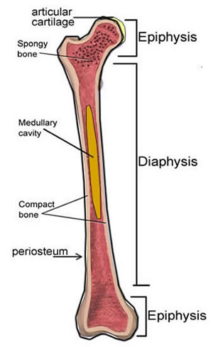
The diaphysis and the epiphysis.
Bone marrow is found in the bone cavities of long bones and is involved in the production of blood cells. Cortical bone appears radiopaque (white) on radiographs as the outermost layer of bone. (see concentric and interstitial lamellae). Let's start by looking at a diagram of bone tissue. Endosteum, centrally, which is derived from derived from a condensation of inner connective tissue, and which helps separate the marrow cavity. Newly formed bone originating from endosteum was observed on day 6. They function in support and movement. Newly formed bone originating from endosteum was observed on day 6. The diaphysis and the epiphysis. Derive their name because they are longer than they are wide. General concepts about skeleton 2. Compact bone that forms the shafts of long bone consists of two structures. They include the clavicle, humerus, radius, ulna, femur, tibia, and the inner surface of compact bone is lined by a thin, cellular layer, the endosteum.
The endosteum appears at the interface between the long bone diagram. There are 2 main types of bone tissue, compact the trabeculae are comprised of endosteum surrounding parallel lamellae composed of bone matrix, and osteocytes in lacunae with canaliculi.


0 Komentar